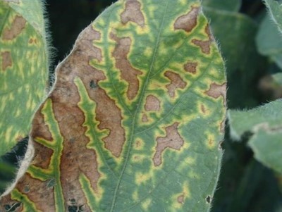
 Interveinal Chlorosis (GROWMARK, Inc.)
- After R3, symptoms of interveinal chlorosis may become evident within the soybean canopy.
- There are many diseases and other issues that could lead to interveinal chlorosis.
- Assessment of field patterns and individual plants is essential.
- Inaccurate identification of the cause of interveinal chlorosis can have negative impacts on future soybean production.
After R3, it is possible to start seeing the canopies of soybeans develop yellow to brown foliage with green veins. This symptom is called interveinal chlorosis. Often this symptom is written off as being due to sudden death syndrome (SDS) in many areas and this year, many are blaming this on red crown rot. However, interveinal chlorosis can be caused by several commonly occurring biotic and abiotic issues. This article will go over some of the most common issues and how to distinguish these issues from one another.
Sudden Death Syndrome (SDS)
SDS is often observed following wet conditions at planting and wet/dry fluctuating conditions after R3. The root infecting pathogen starts to produce toxins that move into the foliage, burning the leaves and causing interveinal chlorosis.
Field pattern: Often low lying or flood prone areas
Plant: When stem is split, the lower stem is necrotic or brown within the pith put the central tissue remains healthy. Presence of blue fungal growth on the base of the stem confirms SDS (Figure 1)

Figure 1. Blue growth on lower stem, which in combination with other symptoms, is indicative of SDS (GROWMARK, Inc.)
Stem Canker
Stem canker is caused by several species of Phomopsis. Some that belong to the Southern stem canker group can produce a toxin that causes interveinal chlorosis. Infection occurs predominantly via windborne spores and infection at the nodes. Eventually stems are colonized resulting in characteristic symptomology.
Field pattern: Wet areas, areas with heavy soybean residue
Plant: Dark lesions that form at nodes and may extend up and down stem (Figure 2). Southern stem canker may cause a lesion to form from crown base up plant, typically up one side of the stem. Often lesions are sunken and framed by black lesion edges. Black, pinhead dots from the fungus may be present within the lesions.

Figure 2. A lesion starting at the node and expanding down the stem. Often Southern stem canker lesions are present on one side of the stem as in this image. (GROWMARK, Inc.)
Brown Stem Rot (BSR)
Brown stem rot is caused by a pathogen that requires soybean residue to persist, and is often observed in continuous soybean production. There are two types of BSR pathogen groups, one that is aggressive and causes interveinal chlorosis, and one that does not cause foliar symptoms. The pathogen infects through the roots during the vegetative stages, eventually rotting out the stem.
Field pattern: wet areas, soybean on soybean systems, cool conditions
Plant: When stem is split, inner portion of the stem will be corky and chocolate brown with a “laddered” look (Figure 3). Dectes stem borer can look similar, but often the larvae will be found within the base of the stem and frass may be present withing the stem or around the stem base.

Figure 3. Brown stem rot causes a brown, corky decay of the inner pith of soybean stems, similar to a brown rot in trees. Decay results in the inner stem becoming chocolate brown in color with a laddered appearance. (GROWMARK, Inc.)
Nematodes
Severe infection by soybean cyst nematode (SCN) and root knot nematode (RKN) can sometimes result in interveinal chlorosis. These sedentary nematodes set up shop on the plant roots, interfering with nutrient and water uptake and pulling nutrients from the plant to sustain nematode growth and development.
Field pattern: sandy areas, dry areas
Plant: Carefully dig out plant and tap off roots. SCN females are present as white, yellow, brown pinhead structures on roots (Figure 4). RKN will cause galling, giving the root an abnormal, often “carrot like” appearance to it.

Figure 4. Small white SCN females on a soybean root. Females are pin head sized and much smaller than nodules. (GROWMARK, Inc.)
Sclerotinia White Mold
White mold is caused by a fungus that launches spores into the air when conditions are cool and wet and soybean canopies have closed. If these spores land on senescing soybean tissues, such as flower petals, they may germinate and grow into stems.
Field pattern: Wet areas, densely canopied areas
Plant: Infected stems are often covered with a white fluffy growth. Black fungal structures resembling rat droppings may be evident on stems and pods, or within stems when split (Figure 5).

Figure 5. Black fungal structures within a soybean stem characteristic of Sclerotinia white mold. (GROWMARK, Inc.)
Red Crown Rot (RCR)
Red crown rot is very similar to SDS in that the fungus infects the roots early when conditions are wet, and then produces a toxin later in the season when conditions alternating between wet and dry after R3. RCR typically is found in warmer climates and areas where peanuts are rotated with soybeans, but the fungus can infect a range of legumes and other plant genera. In addition to the Southern U.S., it is also found in several areas in Illinois, as well as the Midatlantic, and may be present and not diagnosed or misdiagnosed.
Field pattern: Low lying areas. Compacted areas. Poorly drained areas. Warm and wet weather around planting and rain around pod fill.
Plant: Lower stem will be rotted like SDS when split, but inner pith is often grey. Outside of stem and roots contains a red/dark red round fungal growth (Figure 6). Prior to forming these red structures, the stems bases are often very fuzzy and brown. DO NOT assume red discoloration on stem base is RCR! There are many other fungal diseases and saprophytes that can cause a reddish discoloration. Only the presence of perethecia positively confirm this disease in the field. There are also MANY saprophytic species of Fusarium and Microdochium that can grow on decomposing stem tissues. These fungi can become pink with age and may resemble the red perethecia to the untrained eye.

Figure 6. Red crown rot produces red balls on the lower stem. (GROWMARK, Inc.)
Fungicide Burn
Fungicide burn can occur when plants have been growing under cool or cloudy, humid conditions, and there is a sudden change to sunny or hot weather that coincides with a recent fungicide application. Certain fungicides and cultivars are more prone to issues with fungicide burn. In general, products with tebuconazole and prothioconazole tend to be more associated with burn issues when they occur.
Field pattern: Uniform. Symptoms will develop simultaneously, often within a few days of application. New growth should be healthy.
All of these issues have specific management practices that will need to be taken into consideration the next time soybean is planted in that field. However, the practices used depends in the problem at hand. Proper diagnosis of within field issues is essential in strategizing how to attack problematic issues in your fields in the subsequent.
References:
Contact your FS Crop Specialist
Your FS Crop Specialist is here to help you with your nutrient management plans.
Learn more